Lower Body Diagram : Lower Body Diagrams Lzr Ultrabright Powerful Led Therapy / This is the lower part of the stomach.
byAdmin-
0
Lower Body Diagram : Lower Body Diagrams Lzr Ultrabright Powerful Led Therapy / This is the lower part of the stomach.. Labeled illustration chart on white. Abdominal pain and diarrhea (which may be bloody) are symptoms. It contains partially digested food before it. Key bones in the abdominal area include the base of the ribcage and the lumbar spine in the lower back. We hope this picture human body artery diagram in detail can help you study and research.
Anatomynote.com found human body artery diagram in detail from plenty of anatomical pictures on the internet. Jan 21, 2018 · the lower leg is a major anatomical part of the skeletal system. Lower thoracic, lumbar vertebrae and sacrum: An inflammatory condition that usually affects the colon and rectum. Help to prevent injury in many sports that involve the legs.
Sensors Free Full Text Indirect Measurement Of Ground Reaction Forces And Moments By Means Of Wearable Inertial Sensors A Systematic Review Html from www.mdpi.com Key bones in the abdominal area include the base of the ribcage and the lumbar spine in the lower back. Monster walks are a creative way to hit your glutes and hamstrings. The diaphragm forms the upper surface of the abdomen. The muscles of the abdomen protect vital organs underneath and provide structure for the spine. Deep veins, located in the center of the leg near the leg bones, are enclosed by muscle. 2) the tension force t ' 3 exerted by the string on the block. Keeping your back flat and elbows pointed down, push your hips back and lower your body until your thighs are at least parallel to the ground. 1 your spine in this region has a natural inward curve.
Two forces (lower part of figure below) 1) the weight w exerted by the earth on the box.
B) free body diagram of point p; It forms a canal that opens into the vagina, which leads to the outside of the body. Your body organs range from your brain, heart, liver, skin, lungs, kidneys, intestines, stomach, bladder, etc. This is the lower part of the stomach. Jan 21, 2018 · the lower leg is a major anatomical part of the skeletal system. Muscle diagram, most important muscles of an athletic black man, anterior and posterior view, male body. Lower thoracic, lumbar vertebrae and sacrum: Muscle charts of the human body for your reference value these charts show the major superficial and deep muscles of the human body. This curve, called lordosis, helps to: How many muscles are in the back? 1 your spine in this region has a natural inward curve. Veins (in blue) are the blood vessels that return blood to the heart. Female anatomy includes the external genitals, or the vulva, and the internal reproductive organs.
Evenly distribute weights from your upper body into the lower extremities. Food begins breaking down in the body, which is also the largest part of the stomach. Arteries (in red) are the blood vessels that deliver blood to the body. The abdomen (commonly called the belly) is the body space between the thorax (chest) and pelvis. We think this is the most useful anatomy picture that you need.
Blank Human Body Diagram Clip Art Library from clipart-library.com Veins (in blue) are the blood vessels that return blood to the heart. The muscles of the lower back, including the erector spinae and quadratus lumborum muscles, contract to extend and laterally bend the vertebral column. Your lower back (lumbar spine) is the anatomic region between your lowest rib and the upper part of the buttock. Do you ever wonder what the major organs of the body are and. Monster walks are a creative way to hit your glutes and hamstrings. The iliac, femoral, popliteal and tibial (calf) veins are the deep veins in the legs. Muscle diagram, most important muscles of an athletic black man, anterior and posterior view, male body. Three forces (upper part of figure below) 1) tension t 1 2) tension t 2 3) tension t 3
• removing the pelvic lymph nodes • removing the groin lymph nodes • having pelvic and/or groin radiation this pamphlet explains:
It lies between the knee and the ankle, while the upper leg lies between. Arteries (in red) are the blood vessels that deliver blood to the body. Like crohn's disease, bloody diarrhea is a. Deep veins, located in the center of the leg near the leg bones, are enclosed by muscle. The bones of the pelvis and lower back work together to support the body's weight, anchor the abdominal and hip muscles, and protect the delicate vital organs of the vertebral and abdominopelvic cavities. The chapter on the innervation of the lower limb presents diagrams of the lumbosacral plexus and its main nerve branches for the lower limb (lateral cutaneous nerve of the thigh, femoral nerve, sciatic nerve and posterior cutaneous nerve of the thigh and obturator nerve). Key bones in the abdominal area include the base of the ribcage and the lumbar spine in the lower back. The back has a total of 40 muscles. A diagram shows the various inguinal lymph nodes (lymphatic ganglia). In all, there are believed to be 80 organs in your body, all serving different functions and uses. It lies between the knee and the ankle, while the upper leg lies between. Daniel nelson on june 5, 2018 8 comments ! These muscles help the body bend at the waist.
It lies between the knee and the ankle, while the upper leg lies between. Like crohn's disease, bloody diarrhea is a. The lower, narrow part of the uterus (womb) located between the bladder and the rectum. Three forces (upper part of figure below) 1) tension t 1 2) tension t 2 3) tension t 3 Woman holding a blackboard with an illustration of the human digestive system drawn on it in chalk.
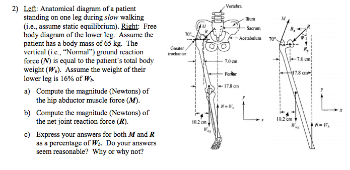
2 Left Anatomical Diagram Of A Patient Standing On Chegg Com from d2vlcm61l7u1fs.cloudfront.net A free body diagram is a graphic, dematerialized, symbolic representation of the body (structure, element or segment of an element) in which all connecting pieces have been removed. Posted on may 24, 2016 by admin. Your lower back (lumbar spine) is the anatomic region between your lowest rib and the upper part of the buttock. The passageway through which fluid passes out of the body during menstrual periods. See here for upper body band. The standard anatomical position is agreed upon by the international medical community. Woman holding a blackboard with an illustration of the human digestive system drawn on it in chalk. In all, there are believed to be 80 organs in your body, all serving different functions and uses.
Keeping your back flat and elbows pointed down, push your hips back and lower your body until your thighs are at least parallel to the ground.
At the level of the pelvic bones, the abdomen. Likewise, there are muscles in other parts of the body that help support and move the spine. The back has a total of 40 muscles. Superficial and deep anterior muscles of upper body The lower, narrow part of the uterus (womb) located between the bladder and the rectum. There are 20 muscle pairs, one on each side of the body. Hold a dumbbell vertically in front of your chest, cupping the top end in both hands. Balance the weight of your head on top of your spine. The vertebral column of the lower back includes the five lumbar vertebrae, the sacrum, and the coccyx. An inflammatory condition that usually affects the colon and rectum. Help to prevent injury in many sports that involve the legs. Female anatomy includes the external genitals, or the vulva, and the internal reproductive organs. Below you'll see diagrams along with the names of the back muscles that may be the cause of your pain.